MRA vs. MRI
When there’s a possibility of having a brain aneurysm, an informed physician will generally request a scan of some sort. Often medical practitioners initially order MRI or CT scan, rather than an MRA. Other practitioners may send the patient home without a scan to treat migraine, stress, or even flu when really there’s an undiagnosed aneurysm. This often results in misdiagnosis leading to potential death or disability from a rupture.
According to research, featured in our Scan2Save campaign, brain aneurysm misdiagnosis occurs in up to one-quarter of patients when seeking initial medical attention.
This blog will look at the distinctions between MRI and MRA scans, how each imaging method is used, and hopefully be a tool in helping save brain aneurysm patients from misdiagnosis and taking their symptoms and health into their own hands. Let’s get started!
What’s an MRI?
Magnetic Resonance Imaging (MRI) uses a gigantic magnet to create detailed images of body tissues, organs, and bones. The MRI machines use radio waves and magnets that bounce off the magnetic field and scan your body parts or tissues.
The generated scan is then sent to a monitor to be viewed and analyzed. At times, radiologists use contrast agents or dyes to gain a clear image of the scanned body part.
Physicians use MRIs to diagnose or rule out a variety of ailments and conditions, including:
- Brain aneurysms
- Arthritis
- Tumors
- Brain injuries
- Cancer
- Inner eye/ear injury
- Torn cartilage, tendons, or ligaments
- Bone infections
- Spinal cord damage
- Multiple sclerosis
Therefore, if you’re experiencing headaches, dizziness, bleeding, mobility problems, etc., an MRI could help diagnose your health problem. As for the aneurysm, the problem may lurk without symptoms. However, if you suspect you have one, an MRI screening can help get a better look and could save your life.
What’s an MRA?
Magnetic resonance angiography (MRA) is a test that helps physicians and radiologists take the blood vessels’ image to diagnose or rule out certain diseases or conditions. In MRA, radio frequency waves and a computer system create detailed images of your body’s major blood vessels, mainly the artery.
Typically, an MRA is performed along with other imaging technologies, including:
- MRI: The non-invasive medical imaging technology discussed above. The MRI and MRA combination can detect brain aneurysms in 60-85% of the cases.
- Fluoroscopy: test whereby the radiologists place a catheter in the blood vessel, often using a contrast dye.
- Computer tomography (CT): This is an imaging test that creates detailed images of the internal body organs, bones, tissues, and blood vessels.
The MRA procedure is used to identify abnormalities in your body, including brain aneurysms and blockages in the blood vessels that could lead to stroke or a heart attack. Usually, the doctor orders an MRA of the head to check the blood vessels leading to the brain.
A doctor may also use MRA to scan for blood clots or identify whether blood vessels have narrowed due to plaque.
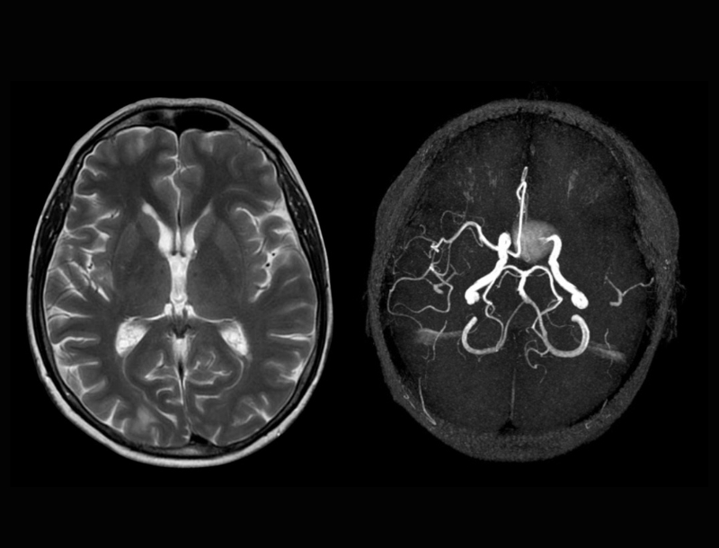
MRA vs. MRI: Similarities and Differences
Both MRA and MRI are non-invasive medical imaging technologies used to diagnose a problem in the inner body. But, the two differ in how they are used to scan.
Here are the key similarities and differences between MRA and MRI.
Similarities:
✔️ Neither procedure use radiation
✔️ They are both non-invasive tools used to diagnose or rule out certain ailments and conditions
Differences:
✔️ Unlike MRA, MRI allows radiologists to examine larger sections of the body
✔️ MRA is primarily meant to diagnose abnormalities in blood vessels, while MRI is used mainly to examine various organs and tissues, including the brain.
The Bottom Line
A brain aneurysm is a weakness in a blood vessel in the brain that balloons and fills with blood. The survival rate for this condition is only about 50%. The survival rate is determined by how early the ailment is diagnosed and treated. By knowing what scans are vital to get when suspecting an aneurysm or showing any symptoms, and advocating for yourself, you could potentially prevent a rupture.
As part of the Scan2Save campaign, this post provides you with information to help you understand the differences between MRA and MRI and how each plays a crucial role in diagnosing brain aneurysms. Misdiagnosis is best prevented by spreading awareness and getting your community involved. Join the Scan2Save campaign can help make a difference and learn more about how the right scan can save your life.
SOURCE USED:
https://bafound.org/diagnosis-and-screening/
https://www.healthimages.com/the-difference-between-an-mri-and-an-mra/