Navigating Imaging Options for Brain Aneurysms

Understanding the various imaging options for detecting and monitoring brain aneurysms is crucial in managing brain health effectively. From less invasive methods like CTA and MRA to the detailed clarity of DSA, each test offers unique strengths, making it vital to choose the one that best fits a patient’s needs. Learn how these imaging methods work, their benefits and limitations, and how they support effective diagnosis and treatment planning
CTA

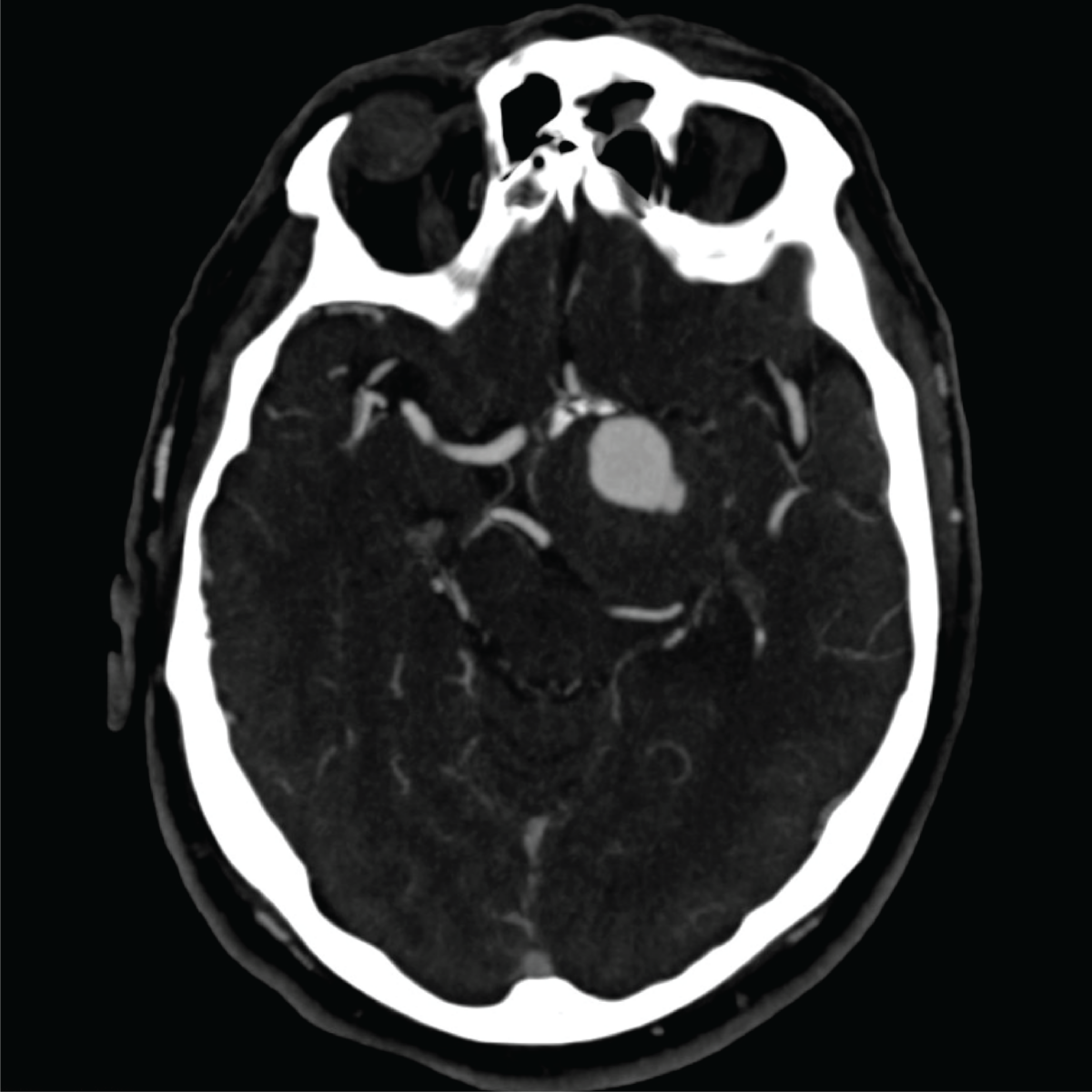
A CT angiogram (CTA) is a type of imaging test that helps doctors examine blood vessels in the brain, particularly to identify aneurysms. Unlike traditional angiograms, CTA is less invasive, involves fewer risks, and provides detailed, high-resolution images that allow doctors to accurately detect and measure larger aneurysms, especially those over 3mm in size. While CTA may not detect very small aneurysms as effectively, it often provides the clarity needed for most cases without exposing patients to as much radiation as other methods. CTA works by combining X-rays with computer processing to create cross-sectional images of the brain. During the procedure, a contrast dye is injected into a vein, which highlights the blood vessels, providing a clear, three-dimensional view of any aneurysms. CTA is especially valuable in emergencies because it is quick, widely accessible, and highly detailed, making it the preferred initial test for diagnosing and assessing aneurysms. For follow-up care, CTA can monitor aneurysms over time, helping ensure that any changes in size or appearance are detected early so that the right care can be provided.

MRA
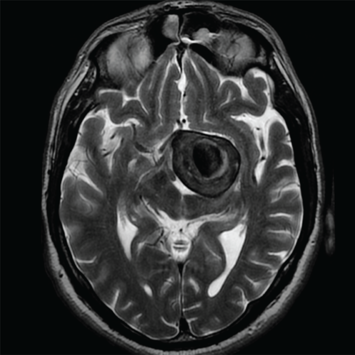
Magnetic resonance angiography (MRA) is a non-invasive imaging test that helps doctors detect brain aneurysms safely, without using radiation or requiring traditional contrast dyes. This makes it especially suitable for regular monitoring, particularly for people with a family history of aneurysms who may need repeated screenings. MRA can accurately detect aneurysms larger than 3mm, though it may be less effective for very small ones. For certain cases where more detailed imaging of an aneurysm’s shape or neck size is needed, doctors may use a type of MRA called contrast-enhanced MRA (CEMRA), which improves visibility and can provide images similar to those from CT angiography. MRA relies on MRI technology, which uses magnetic fields to create detailed images of the body’s soft tissues without radiation. MRA specifically focuses on blood vessels, allowing doctors to observe vascular structures and blood flow patterns in the brain. In some cases, additional MRI sequences, like T1, T2, and FLAIR, can give more insights into surrounding brain tissue or swelling near the aneurysm. While MRI can take longer than a CT scan, it provides high-resolution images and is often preferred for patients who need thorough brain assessments and want to avoid radiation exposure. MRA is also valuable for treatment planning and long-term monitoring of aneurysms, helping doctors maintain a detailed, radiation-free view of the patient’s brain health over time.

DSA
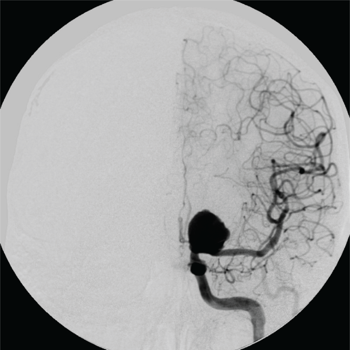
Digital Subtraction Angiography (DSA) is widely regarded as the gold standard for imaging blood vessels in the brain, especially for diagnosing and planning treatments for aneurysms. DSA provides highly detailed images by injecting a contrast dye directly into the bloodstream, allowing doctors to see the precise size, shape, and location of an aneurysm with exceptional clarity. This level of detail is particularly valuable for complex cases and in planning surgical or other treatment approaches. DSA’s advanced version, called 3D DSA, can even reveal aneurysms that may not be visible through other imaging methods. The DSA procedure is more invasive than other imaging tests. It involves threading a small catheter through a blood vessel, usually from the groin or arm, up to the brain, where the dye is released to highlight the blood vessels. While DSA is incredibly accurate, it does carry minor risks, such as a low chance of stroke due to the use of the catheter, and it involves some radiation exposure. However, complications are rare, particularly when performed by experienced specialists. Because of the need for sedation and the small risks involved, DSA is usually reserved for cases where other imaging tests aren’t sufficient or for detailed pre-surgical planning.
Benefits and Drawbacks of the Imaging Techniques
| CTA | MRA | DSA | |
| Advantages | High-resolution images for detecting aneurysms >3mm. Non-invasive compared to DSA. Lower radiation dose compared to DSA. Good for preoperative planning. Can be acquired quickly. Easily accessible. | No radiation exposure Non-invasive and safe for repeated use Contrast-enhanced MRA improves image quality and is comparable to CTA. | Gold standard with the highest accuracy for detecting aneurysms of all sizes. Provides very detailed, 3D images ideal for preoperative planning. Can effectively detect aneurysms near complex anatomical structures. |
| Disadvantages | Uses ionizing radiation. May miss small aneurysms or those near bony structures. Requires iodine-based contrast, which may not be suitable for all patients. Less effective in detecting aneurysms near complex anatomical structures. | May have lower sensitivity for very small aneurysms (<3mm) Less detailed than DSA for complex vascular anatomy. Not appropriate to use in patients with older aneurysm clips and other medical devices. Use of contrast may not be readily available and may be incompatible in some patients (e.g., those with kidney problems). | Invasive procedure with small risk of complications. Uses ionizing radiation and requires iodinated contrast dye. It is time-intensive and carries higher costs due to the need for specialized equipment. Not appropriate for regular monitoring due to invasiveness and radiation exposure. |


 Brain Aneurysm Foundation