

 Brain Aneurysm Foundation
Brain Aneurysm vs. AVM: What’s the Difference?
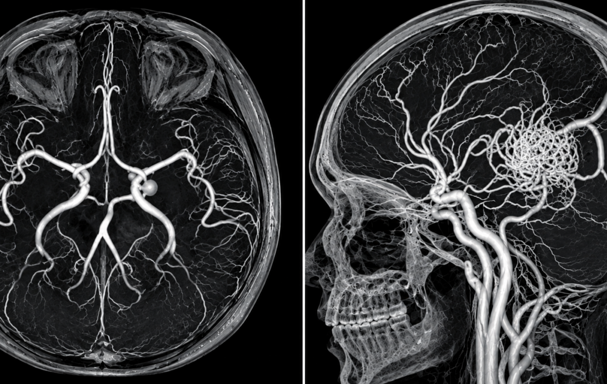
Brain aneurysms and arteriovenous malformations (AVMs) are two distinct conditions that affect the blood vessels in the brain. While both can go undetected and carry a risk of life-threatening bleeding, they differ in how they develop and affect blood flow. Understanding these differences is important for recognizing the risks and the role of early detection and treatment.
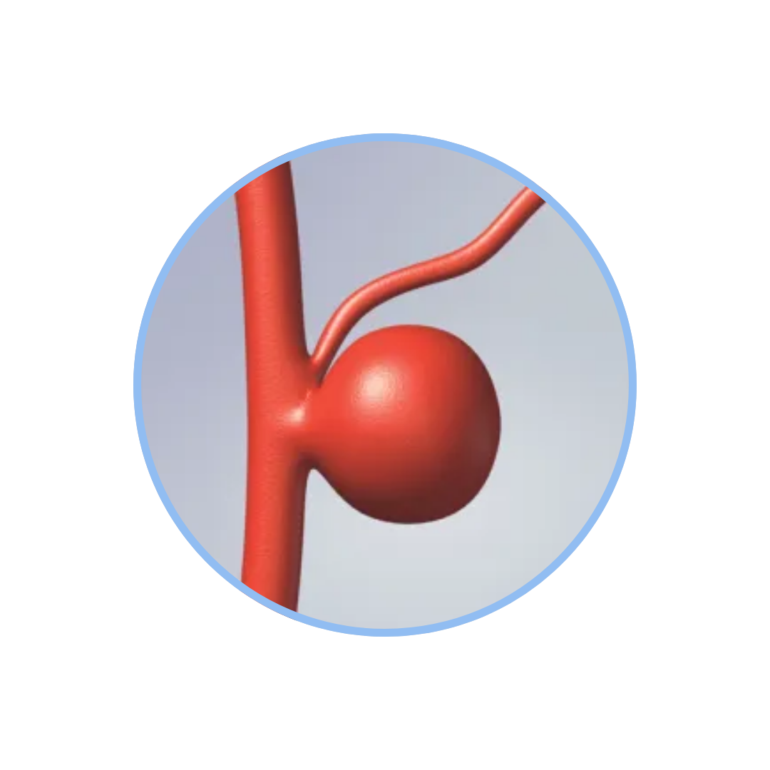
What is a Brain Aneurysm?
A brain aneurysm is a bulge in one of the arteries in or around the brain. They form at weak points in the arteries and are commonly seen where two arteries diverge. Most brain aneurysms go undetected and cause no symptoms. However, if an aneurysm bursts, it is a medical emergency. Ruptured brain aneurysms have a mortality rate of approximately 50%.
What is an Arteriovenous Malformation (AVM)?
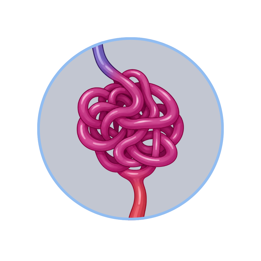
An arteriovenous malformation (AVM) is an abnormal tangle of blood vessels. Typically, blood flows from arteries to capillaries and then to veins. In an AVM, blood flows directly from arteries to veins, bypassing capillaries. This creates a high-flow and high-pressure system that can cause blood vessels to enlarge and weaken. Like aneurysms, there is a potential for AVMs to rupture, but there is a lower mortality rate of 10-15%.
What do they have in common?
- Both aneurysms and AVMs are vascular abnormalities that carry a risk of rupturing.
- Aneurysms can form surrounding or within AVMs as the vessels continue to weaken with high-pressure blood flow.
- They share similar symptoms, including headaches, dizziness, nausea, loss of consciousness, and other neurological deficits.
Symptoms
Brain Aneurysm Symptoms
Unlike AVMs, brain aneurysms usually become symptomatic when they rupture. Common symptoms of a brain aneurysm include:
- sudden and severe headache
- nausea/vomiting
- sensitivity to light
- loss of consciousness
- blurred/double vision.
However, if an aneurysm grows large enough to press on nearby structures, it can cause:
- blurred/double vision
- drooping eyelids
- pain surrounding the eye
- weakness, and numbness
AVM Symptoms
General symptoms of an AVM include:
- sudden/severe headaches
- nausea/vomiting
- seizures.
The location of the AVM can also influence symptoms, as different areas of the brain cause unique symptoms such as:
- dizziness
- poor balance
- vision changes
- difficulty with speech or movement.
These symptoms can develop quickly, but their severity is variable. Additionally, AVMs can become symptomatic before they rupture, allowing for treatment planning with your physician.
Who is at risk of rupture?
Brain Aneurysm Risk Factors and Rupture Risk
- The common risk factors of brain aneurysms include a family history of brain aneurysms, female sex, high blood pressure, smoking, and other genetic diseases (such as polycystic kidney disease).
- In the United States, the annual rate of rupture for brain aneurysms is 8-10 per 100,000 people. While many factors influence how likely an aneurysm is to rupture, the general risk of rupture is approximately 1%.
AVM Risk Factors and Rupture Risk
- AVMs are thought to be congenital, meaning you are born with them. Other risk factors include inheritable diseases, such as hereditary hemorrhagic telangiectasia (HHT), and liver cirrhosis.
- AVMs carry an annual hemorrhage risk of 2-4%, but this risk increases with previous hemorrhages.
Treatment Options
Treatment of Brain Aneurysms
The goal of treating brain aneurysms is to prevent blood flow within the aneurysm sac itself. Many aneurysms are monitored over time by your physician with imaging before treatment options are pursued. If you and your physician decide to treat your aneurysm, this can be accomplished through “clipping”, “coiling”, or “flow diversion”.
- Microsurgical clipping entails an operation where your surgeon will place a small titanium clip at the base, or neck, of the aneurysm. Clipping an aneurysm provides immediate and long-term treatment.
- Endovascular coiling is a procedure where your surgeon can thread a catheter through the arteries from either your wrist or groin to the brain. Once within the arteries of the brain, small platinum coils are placed within the aneurysm itself. This promotes clotting and seals off the aneurysm from the inside.
- Flow diversion is another procedure that is achieved with catheters and treats aneurysms from the inside. In this procedure, a braided wire tube called a stent is placed across the neck of the aneurysm. This stent diverts blood away from the aneurysm itself and stabilizes the artery walls. Over time, the parent blood vessel will grow a new lining over the stent, and the aneurysm will shrink.
Treatment of Arteriovenous Malformations
The goal of AVM treatment is to prevent hemorrhage, and the decision to treat considers patient-associated factors (age, health, symptoms, and other medical conditions) and AVM-associated factors (location, size, and other vascular conditions). There are multiple treatment options, including medical management, surgical resection, stereotactic radiosurgery (SRS), endovascular embolization, or a combination of multiple methods.
- Medical management includes observation and symptom control, such as anti-seizure medications.
- Surgical resection entails an operation to remove the tangle of blood vessels under general anesthesia.
- SRS is a non-invasive method that uses focused radiation beams to shrink the AVM over the course of 2-3 years. Endovascular embolization is a minimally invasive procedure that delivers materials to the AVM by a catheter to plug the lesion.
- Embolization may be used in combination with surgical resection and SRS to reduce the size of the AVM and optimize the effectiveness of treatment.