

 Brain Aneurysm Foundation
 Brain Aneurysm Imaging and Treatment: Advances That Offer New Hope
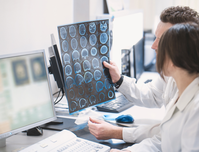
When telling patients – and their loved ones – about a brain aneurysm, specialists are often met with concern. Then the questions come: How serious is it? Could it rupture? Does it need treatment? What do I do now?
Fortunately, advances in brain aneurysm imaging and minimally invasive treatment are giving specialists more information and more options to personalize care. That’s been helpful for the 1 in 50 people – an estimated 6.8 million Americans – living with an unruptured brain aneurysm.
Advances in Imaging: Seeing More Than the Aneurysm
Two important non-invasive imaging tests used to evaluate brain aneurysms are CT angiography of the head and neck (CTA H/N) and magnetic resonance angiography (MRA) of the brain.
CTA uses computed tomography and contrast material to create detailed images of blood vessels. Because it is fast and widely available, CTA is particularly useful when doctors suspect a brain aneurysm during an emergency. MRA uses magnetic resonance imaging to visualize blood vessels without the radiation associated with CT.
Research shows that both CTA and MRA provide effective diagnostic information for detecting intracranial aneurysms, but very small aneurysms and certain areas of the head and neck can be more challenging to evaluate.
Looking at the Aneurysm Wall
Newer technology is high-resolution MRA with vessel wall imaging (MR-VWI). Rather than looking only at the shape and blood flow within an aneurysm, this tool can provide information about the aneurysm wall itself.
Following contrast administration, this imaging can show areas where the aneurysm wall enhances, which is a marker for ongoing inflammation predicting instability, future growth, and rupture. While promising, as noted in a recent study, vessel wall imaging is considered another piece of information rather than a definitive prediction that an aneurysm will rupture.
DSA: The Definitive Imaging Test
Even with advances in non-invasive imaging, digital subtraction angiography (DSA) remains the definitive imaging test for cerebral aneurysms.
DSA is a catheter-based procedure that provides highly detailed, real-time images of blood vessels and aneurysms. It can show the precise anatomy needed to determine whether treatment is appropriate and to plan an endovascular procedure.
Because DSA is invasive, it carries procedural risks. The decision to perform DSA is therefore individualized based on the information needed to safely manage the aneurysm.
Minimally Invasive Treatment Options
With a doctor’s recommendation, many aneurysms can now be treated from inside the blood vessels, avoiding traditional open brain surgery.
Primary Coiling
with or without stenting
With primary coiling, tiny platinum coils are placed inside the aneurysm to promote blood stasis and clot formation while protecting the primary artery. For some wide-neck aneurysms, a stent can be placed across the opening to help keep the coils in position and protect the primary artery.
New research supports stent-assisted coiling as an effective treatment option for appropriately selected aneurysms.
Flow-Diverting
Stents
A flow-diverting stent is placed across the aneurysm’s opening to redirect blood flow away from the aneurysm. This promotes stasis and clotting inside the aneurysm while encouraging the vessel lining to grow across the device, a process known as reendothelialization.
Studies have demonstrated high rates of aneurysm occlusion with modern flow-diversion technology, although patient selection and antiplatelet therapy are important considerations.
Intrasaccular
Devices
An intrasaccular device is placed directly inside the aneurysm rather than in the primary artery. The device disrupts blood flow at the aneurysm’s base or neck, preventing blood from entering the aneurysm and promoting thrombosis.
Long-term research results demonstrate adequate aneurysm occlusion in over 87% of patients at five years, supporting its role as an option for appropriately selected aneurysms.
A More Personalized Path Forward
For patients and families, perhaps the most encouraging advance is not one scan or device, but the ability to combine increasingly detailed imaging with multiple treatment options.
Not every brain aneurysm needs to be treated. Some can be safely monitored, while others may benefit from intervention based on factors such as size, location, shape, growth, symptoms and other individual risk factors.
Today, specialists have more ways to see, understand and treat brain aneurysms than ever before. If you or someone you love has been diagnosed with a brain aneurysm, talk with a cerebrovascular or neuro-interventional specialist about your imaging findings and the treatment or monitoring plan that is right for you.
Joshua Santucci, MD is a Vascular and Neuro-interventional Neurologist at Endeavor Health Neurosciences Institute in the Chicago area.